APD-III
* Please be kindly noted products are not for therapeutic use. We do not sell to patients.
| Category | Antibiotics |
| Catalog number | BBF-03289 |
| CAS | 87098-48-4 |
| Molecular Weight | 1036.34 |
| Molecular Formula | C53H93N7O13 |
Online Inquiry
Description
APD-III is an ester peptide antibiotic produced by Bacillus subtilis C-756. It can inhibit the activity of cyclic-AMP phosphodiesterase.
Properties
| Appearance | Powder |
| Melting Point | 139-140°C |
Reference Reading
1. Seminal vesicles and diabetic neuropathy: ultrasound evaluation after prolonged treatment with a selective phosphodiesterase-5 inhibitor
S La Vignera, R A Condorelli, E Vicari, F Lotti, V Favilla, G Morgia, M Maggi, A E Calogero Andrology. 2013 Mar;1(2):245-50. doi: 10.1111/j.2047-2927.2012.00025.x. Epub 2012 Nov 29.
We have previously reported that infertile patients with diabetes mellitus (DM) have a particular ultrasound features of the seminal vesicles (SV) characterized by higher fundus-to-body ratio and lower pre- and post-ejaculatory difference in body antero-posterior diameter (APD). Based on these premises the aim of the present study was to investigate possible ultrasound SV changes in infertile patients with DM and diabetic neuropathy (DN), after prolonged administration of tadalafil (TAD) (a specific phosphodiesterase-5 inhibitor). To accomplish this, 20 infertile patients with symptomatic DN and erectile dysfunction were selected and arbitrarily divided into two groups which were assigned to: daily administration of 5 mg TAD for 3 months (Group A) (n = 10) and administration of placebo (Group B) (n = 10). All patients underwent to scrotal and prostate-vesicular transrectal ultrasound evaluation and semen analysis (Laboratory Manual for the Examination and Processing of Human Semen, WHO, 2010) before and after treatment. The following SV US parameters were recorded: (i) body APD; (ii) fundus APD; (iii) parietal thickness of the right and left SVs; and (iv) number of polycyclic areas within both SVs. We then calculated the following parameters: (i) fundus/body (F/B) ratio; (ii) difference of the parietal thickness between the right and the left SV and (iii) pre- and post-ejaculatory APD difference. In addition, we also evaluated the SV ejection fraction. Group A patients showed a significant reduction in F/B ratio and higher pre- and post-ejaculatory body SV APD difference compared with baseline or Group B after 3 months. These patients showed also a significant increase in SV ejection fraction and a significant improvement of the total sperm count, progressive motility, seminal levels of fructose, leucocytes and ejaculate volume. In conclusion, these results suggest that infertile DM patients with DN and erectile dysfunction had an improvement of ultrasound features suggestive of diabetic neuropathy after daily treatment with low doses of TAD.
2. Acylpeptides, the inhibitors of cyclic adenosine 3',5'-monophosphate phosphodiesterase. I. Purification, physicochemical properties and structures of fatty acid residues
K Hosono, H Suzuki J Antibiot (Tokyo). 1983 Jun;36(6):667-73. doi: 10.7164/antibiotics.36.667.
An inhibitor of cyclic adenosine 3',5'-monophosphate (cAMP) phosphodiesterase was isolated from the culture filtrate of Bacillus subtilis C-756 isolated from soil. It was purified and finally separated into three fractions by reverse-phase HPLC. The respective fractions were designated as APD-I, -II and -III in the order eluted and the relative quantities of APD-I, -II and -III were approximately 10%, 40% and 50%, respectively. They were acylpeptides composed of beta-hydroxy fatty acid residues and heptapeptide. Though the amino acid compositions of the peptides were the same, the fatty acid residues were all different. APD-I contained a mixture of 3-hydroxy-11-methyldodecanoic acid (i-C13h3) and 3-hydroxy-10-methyldodecanoic acid (a-C13h3). APD-II contained 3-hydroxytetradecanoic acid (n-C14h3). APD-III contained a mixture of 3-hydroxy-13-methyltetradecanoic acid (i-C15h3) and 3-hydroxy-12-methyltetradecanoic acid (a-C15h3).
3. Seminal vesicles and diabetic neuropathy: ultrasound evaluation in patients with couple infertility and different levels of glycaemic control
Sandro La Vignera, Rosita A Condorelli, Enzo Vicari, Rosario D'Agata, Aldo E Calogero Asian J Androl. 2011 Nov;13(6):872-6. doi: 10.1038/aja.2011.47. Epub 2011 Aug 1.
The aim of this study was to evaluate the ultrasound characteristics of the seminal vesicles (SVs) of infertile patients with diabetes mellitus (DM) and diabetic neuropathy (DN) and to investigate possible changes in ultrasound characteristics related to glycaemic control. To accomplish this, 45 infertile patients with type 2 DM and symptomatic DN were selected. Twenty healthy fertile men and 20 patients with idiopathic oligoasthenoteratozoospermia without DM represented the control groups. DM patients were arbitrarily divided into three groups according to glycaemic control level (A=glycosylated haemoglobin 10%). Patients underwent prostate-vesicular transrectal ultrasonography and sperm analysis. The following SV ultrasound parameters were recorded: (i) body antero-posterior diameter (APD); (ii) fundus APD; (iii) parietal thicknesses of the right and left SVs; and (iv) the number of polycyclic areas within both SVs. We then calculated the following parameters: (i) fundus/body (F/B) ratio; (ii) difference of the parietal thickness between the right and the left SV; and (iii) pre- and post-ejaculatory APD difference. All DM patients had a higher F/B ratio compared to controls (P<0.05). Group C had a higher F/B ratio compared to the other DM groups (P<0.05). All DM patients had a lower pre- and post-ejaculatory difference of the body SV APD compared to controls (P<0.05). Groups A and B had a similar pre- and post-ejaculatory difference of the body SV APD, whereas this difference was lower in Group C (P<0.05). In conclusion, infertile DM patients with DN showed peculiar SV ultrasound features suggestive of functional atony, and low glycaemic control was associated with greater expression of these features.
Recommended Products
| BBF-02614 | Nystatin | Inquiry |
| BBF-01829 | Deoxynojirimycin | Inquiry |
| BBF-00677 | 3-Amino-3-deoxy-D-glucose | Inquiry |
| BBF-00764 | Cerebroside C | Inquiry |
| BBF-03753 | Baicalin | Inquiry |
| BBF-01826 | Deoxymannojirimycin | Inquiry |
Bio Calculators
* Our calculator is based on the following equation:
Concentration (start) x Volume (start) = Concentration (final) x Volume (final)
It is commonly abbreviated as: C1V1 = C2V2
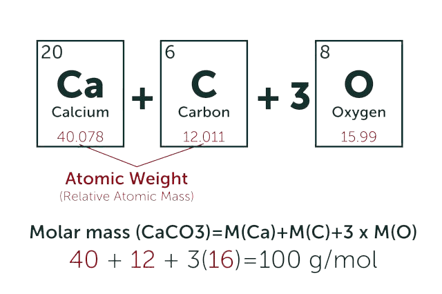
* Total Molecular Weight:
g/mol
Tip: Chemical formula is case sensitive. C22H30N4O √ c22h30n40 ╳