Avilamycin A
* Please be kindly noted products are not for therapeutic use. We do not sell to patients.

| Category | Antibiotics |
| Catalog number | BBF-00233 |
| CAS | 69787-79-7 |
| Molecular Weight | 1404.23 |
| Molecular Formula | C61H88Cl2O32 |
Online Inquiry
Description
Avilamycin A is produced by the strain of Streptomyces viridochromogenes NRRL 2860. It inhibits the binding of amino acid-based tRNA to the bacterial ribosome for 30S subunit and has anti-gram-positive bacterial activity. It has been tried as feed additive in animal feeding.
Specification
| Synonyms | Flambamycin, 23-deoxy- |
| IUPAC Name | [(2R,3S,4R,6S)-6-[(2'R,3'S,3aR,4R,4'R,6S,7aR)-6-[(2S,3R,4R,5S,6R)-2-[(2R,3S,4S,5S,6S)-6-[(2R,3aS,3'aR,6S,6'R,7R,7'S,7aR,7'aR)-7'-acetyl-7'-hydroxy-6'-methyl-7-(2-methylpropanoyloxy)spiro[4,6,7,7a-tetrahydro-3aH-[1,3]dioxolo[4,5-c]pyran-2,4'-6,7a-dihydro-3aH-[1,3]dioxolo[4,5-c]pyran]-6-yl]oxy-4-hydroxy-5-methoxy-2-(methoxymethyl)oxan-3-yl]oxy-3-hydroxy-5-methoxy-6-methyloxan-4-yl]oxy-4'-hydroxy-2',4,7a-trimethylspiro[3a,4,6,7-tetrahydro-[1,3]dioxolo[4,5-c]pyran-2,6'-oxane]-3'-yl]oxy-4-hydroxy-2-methyloxan-3-yl] 3,5-dichloro-4-hydroxy-2-methoxy-6-methylbenzoate |
| Canonical SMILES | CC1C(C(CC(O1)OC2C(OC3(CC2O)OC4C(OC(CC4(O3)C)OC5C(C(OC(C5OC)C)OC6C(OC(C(C6O)OC)OC7C(C8C(CO7)OC9(O8)C1C(C(C(O9)C)(C(=O)C)O)OCO1)OC(=O)C(C)C)COC)O)C)C)O)OC(=O)C1=C(C(=C(C(=C1OC)Cl)O)Cl)C |
| InChI | InChI=1S/C61H88Cl2O32/c1-21(2)53(70)87-49-45-32(92-61(93-45)52-51(78-20-79-52)60(72,27(8)64)28(9)91-61)19-77-56(49)89-57-48(76-14)39(68)44(31(83-57)18-73-11)88-55-40(69)47(43(74-12)24(5)82-55)85-34-17-58(10)50(26(7)81-34)94-59(95-58)16-30(66)42(25(6)90-59)84-33-15-29(65)41(23(4)80-33)86-54(71)35-22(3)36(62)38(67)37(63)46(35)75-13/h21,23-26,28-34,39-45,47-52,55-57,65-69,72H,15-20H2,1-14H3/t23-,24-,25-,26-,28-,29-,30-,31-,32+,33+,34+,39+,40-,41-,42-,43+,44-,45-,47-,48+,49-,50-,51-,52-,55+,56+,57+,58-,59?,60+,61-/m1/s1 |
| InChI Key | XIRGHRXBGGPPKY-FCNCREMHSA-N |
Properties
| Appearance | Colorless Acicular Crystalline |
| Antibiotic Activity Spectrum | gram-positive bacterial |
| Melting Point | 188-189.5 °C |
| Solubility | Soluble in ethyl acetate, benzene, chloroform and ethyl acetate. |
Reference Reading
1. Analysis of a C-methyltransferase gene (aviG1) involved in avilamycin biosynthesis in Streptomyces viridochromogenes Tü57 and complementation of a Saccharopolyspora erythraea eryBIII mutant by aviG1
G Weitnauer, S Gaisser, L Kellenberger, P F Leadlay, A Bechthold Microbiology (Reading) . 2002 Feb;148(Pt 2):373-379. doi: 10.1099/00221287-148-2-373.
Streptomyces viridochromogenes Tü57 is the principal producer of avilamycin A. aviG1, a putative methyltransferase gene, was detected in the avilamycin biosynthetic gene cluster. To determine the function of aviG1, a targeted gene inactivation experiment was performed. The resulting chromosomal mutant, carrying an in-frame deletion in aviG1, was deficient in avilamycin production. aviG1 was used to complement an eryBIII mutant of the erythromycin A producer Saccharopolyspora erythraea [Gaisser, S., Bohm, G. A., Doumith, M., Raynal, M. C., Dhillon, N., Cortes, J. & Leadlay, P. F. (1998). Mol Gen Genet 258, 78-88]. The presence of erythromycin A in the culture supernatant of the complemented mutant indicated that L-mycarose biosynthesis could be restored and that AviG1 could take over the function of the C-methyltransferase EryBIII.
2. Fragmentation studies on the antibiotic avilamycin A using ion trap mass spectrometry
Diana S Aga, Sandra Pérez, Peter Eichhorn, Andreas Bechtholt J Mass Spectrom . 2004 Dec;39(12):1541-53. doi: 10.1002/jms.760.
A comprehensive study on the fragmentation pattern of the antimicrobial growth promoter avilamycin A was conducted in a quadrupole ion trap mass spectrometer equipped with an electrospray ionization (ESI) source. Performing multiple-stage experiments on the deprotonated molecule (m/z 1401) and its principal product ions showed that a sequential shortening of the oligosaccharide backbone took place, which can be attributed to the localization of the negative charge in the terminal dichloroisoeverninic acid. Under (+)-ESI conditions, avilamycin A readily formed an intense sodium-cationized molecule, [M + Na](+) (m/z 1425). Structural elucidation of the second-, third- and fourth-generation fragment ions revealed that all of the structures shared a common molecular portion comprising a central monosaccharide. This observation allowed us to assign confidently the complexation site of the alkali metal cation. In addition to the monosodiated molecule, the full-scan mass spectral acquisition also yielded a less abundant disodiated molecule, [M - H + 2Na](+) (m/z 1447). Multiple-stage experiments on this ion indicated that the second sodium ion compensates for the negative charge located at either of two positions within the molecule. While deprotonation of the phenolic hydroxyl group in the dichloroisoeverninic acid moiety was suggested to be driven by charge stabilization in the aromatic ring (in analogy with the deprotonated molecule in the (-)-ESI mode), the deprotonation at an alpha-carbon of an ester side-chain substituent in the oligosaccharide part was believed to provide a stable chelation-like coordination site for the cation.
3. Determination of total avilamycin residues as dichloroisoeverninic acid in porcine muscle, fat, and liver by LC-MS/MS
Satoru Nemoto, Shizuka Saito-Shida, Tomoko Hayashi, Hiroshi Akiyama Food Chem . 2018 May 30;249:84-90. doi: 10.1016/j.foodchem.2018.01.003.
A sensitive and reliable method for determining the total avilamycin residues was developed using LC-MS/MS. Avilamycin (consisting of avilamycin A and 15 other minor factors) and its metabolites in porcine muscle, fat, and liver were analysed as the marker residue dichloroisoeverninic acid (DIA), in accordance with the maximum residue limit (MRL) established by international organisations such as Codex Alimentarius Commission and other regulatory bodies. The analytes were extracted from samples with acetone, hydrolysed to DIA, partitioned into ethyl acetate, and cleaned up prior to the LC-MS/MS analysis. The method was validated at Codex MRL and 0.01 mg/kg. The results show excellent recoveries ranging from 100 to 108%, with the relative standard deviations <6%. Matrix effects were negligible for all types of samples, indicating effective sample clean-up. The absence of interfering peaks close to the retention time in blank samples demonstrates high selectivity. Overall, this method is reliable and suitable for regulatory-purpose analysis.
Recommended Products
| BBF-02642 | Lactonamycin | Inquiry |
| BBF-03753 | Baicalin | Inquiry |
| BBF-03754 | CASTANOSPERMINE | Inquiry |
| BBF-01826 | Deoxymannojirimycin | Inquiry |
| BBF-01829 | Deoxynojirimycin | Inquiry |
| BBF-00764 | Cerebroside C | Inquiry |
Bio Calculators
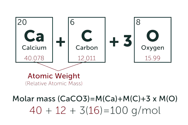
* Our calculator is based on the following equation:
Concentration (start) x Volume (start) = Concentration (final) x Volume (final)
It is commonly abbreviated as: C1V1 = C2V2
* Total Molecular Weight:
g/mol
Tip: Chemical formula is case sensitive. C22H30N4O √ c22h30n40 ╳