Mycinamicin IV
* Please be kindly noted products are not for therapeutic use. We do not sell to patients.
| Category | Antibiotics |
| Catalog number | BBF-01969 |
| CAS | |
| Molecular Weight | 695.88 |
| Molecular Formula | C37H61NO11 |
Online Inquiry
Description
Mycinamicin IV is a macrolide antibiotic produced by Micromonospora griseorubida sp. nov. Activity against gram-positive bacteria.
Specification
| Synonyms | 12,13-Deepoxy-12,13-didehydromycinamicin I |
| IUPAC Name | (3E,5S,6S,7S,9R,11E,13E,15R,16R)-6-[(2S,3R,4S,6R)-4-(dimethylamino)-3-hydroxy-6-methyloxan-2-yl]oxy-16-ethyl-15-[[(2R,3R,4R,5R,6R)-5-hydroxy-3,4-dimethoxy-6-methyloxan-2-yl]oxymethyl]-5,7,9-trimethyl-1-oxacyclohexadeca-3,11,13-triene-2,10-dione |
| Canonical SMILES | CCC1C(C=CC=CC(=O)C(CC(C(C(C=CC(=O)O1)C)OC2C(C(CC(O2)C)N(C)C)O)C)C)COC3C(C(C(C(O3)C)O)OC)OC |
| InChI | InChI=1S/C37H61NO11/c1-11-29-26(20-45-37-35(44-10)34(43-9)31(41)25(6)47-37)14-12-13-15-28(39)22(3)18-23(4)33(21(2)16-17-30(40)48-29)49-36-32(42)27(38(7)8)19-24(5)46-36/h12-17,21-27,29,31-37,41-42H,11,18-20H2,1-10H3/b14-12+,15-13+,17-16+/t21-,22+,23-,24+,25+,26+,27-,29+,31+,32+,33+,34+,35+,36-,37+/m0/s1 |
| InChI Key | DBTIHDIIXPQOFR-JMHKOBKLSA-N |
Properties
| Antibiotic Activity Spectrum | Gram-positive bacteria |
| Boiling Point | 801.3±65.0°C at 760 mmHg |
| Melting Point | 174-176°C |
| Density | 1.2±0.1 g/cm3 |
Reference Reading
1. Total Synthesis of Mycinamicin IV as Integral Part of a Collective Approach to Macrolide Antibiotics
Georg Späth, Alois Fürstner Chemistry. 2022 Feb 19;28(11):e202104400. doi: 10.1002/chem.202104400. Epub 2022 Jan 10.
The total synthesis of the 16-membered macrolide mycinamicin IV is outlined, which complements our previously disclosed, largely catalysis-based route to the aglycone. This work must also be seen in the context of our recent conquest of aldgamycin N, a related antibiotic featuring a similar core but a distinctly different functionalization pattern. Taken together, these projects prove that the underlying blueprint is integrative and hence qualifies for a collective approach to this prominent class of natural products. In both cases, the final glycosylation phase mandated close attention and was accomplished only after robust de novo syntheses of the (di)deoxy sugars of the desosamine, chalcose, mycinose and aldgarose types had been established. Systematic screening of the glycosidation promoter was also critically important for success.
2. Substrate recognition by two different P450s: Evidence for conserved roles in a common fold
Drew R Tietz, Allison M Colthart, Susan Sondej Pochapsky, Thomas C Pochapsky Sci Rep. 2017 Oct 19;7(1):13581. doi: 10.1038/s41598-017-14011-w.
Cytochrome P450 monooxygenases CYP101A1 and MycG catalyze regio- and stereospecific oxidations of their respective substrates, d-camphor and mycinamicin IV. Despite the low sequence homology between the two enzymes (29% identity) and differences in size and hydrophobicity of their substrates, the conformational changes that occur upon substrate binding in both enzymes as determined by solution NMR methods show some striking similarities. Many of the same secondary structural features in both enzymes are perturbed, suggesting the existence of a common mechanism for substrate binding and recognition in the P450 superfamily.
3. Solution Conformations and Dynamics of Substrate-Bound Cytochrome P450 MycG
Drew R Tietz, Larissa M Podust, David H Sherman, Thomas C Pochapsky Biochemistry. 2017 May 30;56(21):2701-2714. doi: 10.1021/acs.biochem.7b00291. Epub 2017 May 16.
MycG is a P450 monooxygenase that catalyzes the sequential hydroxylation and epoxidation of mycinamicin IV (M-IV), the last two steps in the biosynthesis of mycinamicin II, a macrolide antibiotic isolated from Micromonospora griseorubida. The crystal structure of MycG with M-IV bound was previously determined but showed the bound substrate in an orientation that did not rationalize the observed regiochemistry of M-IV hydroxylation. Nuclear magnetic resonance paramagnetic relaxation enhancements provided evidence of an orientation of M-IV in the MycG active site more compatible with the observed chemistry, but substrate-induced changes in the enzyme structure were not characterized. We now describe the use of amide 1H-15N residual dipolar couplings as experimental restraints in solvated "soft annealing" molecular dynamics simulations to generate solution structural ensembles of M-IV-bound MycG. Chemical shift perturbations, hydrogen-deuterium exchange, and 15N relaxation behavior provide insight into the dynamic and electronic perturbations in the MycG structure in response to M-IV binding. The solution and crystallographic structures are compared, and the possibility that the crystallographic orientation of bound M-IV represents an inhibitory mode is discussed.
Recommended Products
| BBF-01826 | Deoxymannojirimycin | Inquiry |
| BBF-00764 | Cerebroside C | Inquiry |
| BBF-02614 | Nystatin | Inquiry |
| BBF-05862 | Epirubicin | Inquiry |
| BBF-01693 | Doxorubicin EP Impurity A (Daunorubicin) | Inquiry |
| BBF-03755 | Actinomycin D | Inquiry |
Bio Calculators
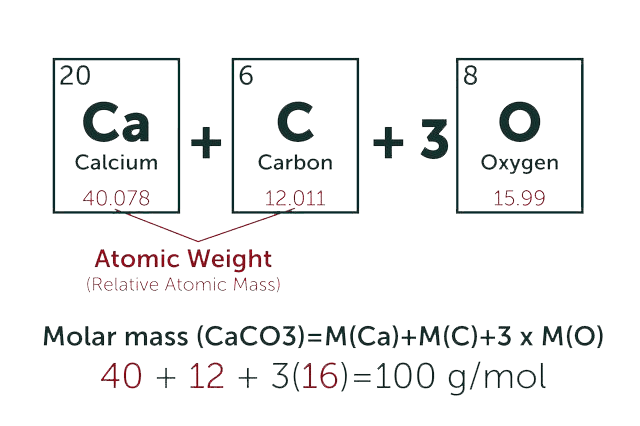
* Our calculator is based on the following equation:
Concentration (start) x Volume (start) = Concentration (final) x Volume (final)
It is commonly abbreviated as: C1V1 = C2V2
* Total Molecular Weight:
g/mol
Tip: Chemical formula is case sensitive. C22H30N4O √ c22h30n40 ╳