N-Acetyl-L-prolinol
* Please be kindly noted products are not for therapeutic use. We do not sell to patients.

| Category | Others |
| Catalog number | BBF-05189 |
| CAS | 66158-68-7 |
| Molecular Weight | 143.18 |
| Molecular Formula | C7H13NO2 |
| Purity | >95% by HPLC |
Online Inquiry
Specification
| Related CAS | 123958-87-2 (D-configuration) |
| Synonyms | 1-((S)-2-Hydroxymethyl-pyrrolidin-1-yl)-ethanone; acetylprolinol; Ethanone, 1-[(2S)-2-(hydroxymethyl)-1-pyrrolidinyl]-; (S)-1-(2-(hydroxymethyl)pyrrolidin-1-yl)ethanone; (S)-1-acetyl-2-(hydroxymethyl)pyrrolidine; (S)-N-acetylpyrrolidinylmethanol; (S)-N-acetylprolinol |
| Storage | Store at -20°C |
| IUPAC Name | 1-[(2S)-2-(hydroxymethyl)pyrrolidin-1-yl]ethanone |
| Canonical SMILES | CC(=O)N1CCCC1CO |
| InChI | InChI=1S/C7H13NO2/c1-6(10)8-4-2-3-7(8)5-9/h7,9H,2-5H2,1H3/t7-/m0/s1 |
| InChI Key | VQNVOVAHJAHGQP-ZETCQYMHSA-N |
Properties
| Boiling Point | 295.2±13.0°C at 760 mmHg |
| Density | 1.1±0.1 g/cm3 |
Reference Reading
1. Unconventional N-H…N Hydrogen Bonds Involving Proline Backbone Nitrogen in Protein Structures
R N V Krishna Deepak, Ramasubbu Sankararamakrishnan Biophys J. 2016 May 10;110(9):1967-79. doi: 10.1016/j.bpj.2016.03.034.
Contrary to DNA double-helical structures, hydrogen bonds (H-bonds) involving nitrogen as the acceptor are not common in protein structures. We systematically searched N-H…N H-bonds in two different sets of protein structures. Data set I consists of neutron diffraction and ultrahigh-resolution x-ray structures (0.9 Å resolution or better) and the hydrogen atom positions in these structures were determined experimentally. Data set II contains structures determined using x-ray diffraction (resolution ≤ 1.8 Å) and the positions of hydrogen atoms were generated using a computational method. We identified 114 and 14,347 potential N-H…N H-bonds from these two data sets, respectively, and 56-66% of these were of the Ni+1-Hi+1…Ni type, with Ni being the proline backbone nitrogen. To further understand the nature of such unusual contacts, we performed quantum chemical calculations on the model compound N-acetyl-L-proline-N-methylamide (Ace-Pro-NMe) with coordinates taken from the experimentally determined structures. A potential energy profile generated by varying the ψ dihedral angle in Ace-Pro-NMe indicates that the conformation with the N-H…N H-bond is the most stable. An analysis of H-bond-forming proline residues reveals that more than 30% of the proline carbonyl groups are also involved in n → π(*) interactions with the carbonyl carbon of the preceding residue. Natural bond orbital analyses demonstrate that the strength of N-H…N H-bonds is less than half of that observed for a conventional H-bond. This study clearly establishes the H-bonding capability of proline nitrogen and its prevalence in protein structures. We found many proteins with multiple instances of H-bond-forming prolines. With more than 15% of all proline residues participating in N-H…N H-bonds, we suggest a new, to our knowledge, structural role for proline in providing stability to loops and capping regions of secondary structures in proteins.
2. Experimental conformational energy maps of proteins and peptides
Govardhan A Balaji, H G Nagendra, Vitukudi N Balaji, Shashidhar N Rao Proteins. 2017 Jun;85(6):979-1001. doi: 10.1002/prot.25266. Epub 2017 Mar 7.
We have presented an extensive analysis of the peptide backbone dihedral angles in the PDB structures and computed experimental Ramachandran plots for their distributions seen under a various constraints on X-ray resolution, representativeness at different sequence identity percentages, and hydrogen bonding distances. These experimental distributions have been converted into isoenergy contour plots using the approach employed previously by F. M. Pohl. This has led to the identification of energetically favored minima in the Ramachandran (ϕ, ψ) plots in which global minima are predominantly observed either in the right-handed α-helical or the polyproline II regions. Further, we have identified low energy pathways for transitions between various minima in the (ϕ,ψ) plots. We have compared and presented the experimental plots with published theoretical plots obtained from both molecular mechanics and quantum mechanical approaches. In addition, we have developed and employed a root mean square deviation (RMSD) metric for isoenergy contours in various ranges, as a measure (in kcal.mol-1 ) to compare any two plots and determine the extent of correlation and similarity between their isoenergy contours. In general, we observe a greater degree of compatibility with experimental plots for energy maps obtained from molecular mechanics methods compared to most quantum mechanical methods. The experimental energy plots we have investigated could be helpful in refining protein structures obtained from X-ray, NMR, and electron microscopy and in refining force field parameters to enable simulations of peptide and protein structures that have higher degree of consistency with experiments. Proteins 2017; 85:979-1001. © 2017 Wiley Periodicals, Inc.
3. Identification, characterization, and cloning of a novel aminoacylase, L-pipecolic acid acylase from Pseudomonas species
Junji Hayashi, Yoshiaki Ichiki, Akiko Kanda, Kazuyoshi Takagi, Mamoru Wakayama J Gen Appl Microbiol. 2021 Nov 25;67(5):186-194. doi: 10.2323/jgam.2020.12.001. Epub 2021 Jun 26.
L-Pipecolic acid is utilized as a vital component of specific chemical compounds, such as immunosuppressive drugs, anticancer reagents, and anesthetic reagents. We isolated and characterized a novel L-aminoacylase, N-acetyl-L-pipecolic acid-specific aminoacylase (LpipACY), from Pseudomonas sp. AK2. The subunit molecular mass of LpipACY was 45 kDa and was assumed to be a homooctamer in solution. The enzyme exhibited high substrate specificity toward N-acetyl-L-pipecolic acid and a high activity for N-acetyl-L-pipecolic acid and N-acetyl-L-proline. This enzyme was stable at a high temperature (60°C for 10 min) and under an alkaline pH (6.0-11.5). The N-terminal and internal amino acid sequences of the purified enzyme were STTANTLILRNG and IMASGGV, respectively. These sequences are highly consistent with those of uncharacterized proteins from Pseudomonas species, such as amidohydrolase and peptidase. We also cloned and overexpressed the gene coding LpipACY in Escherichia coli. Moreover, the recombinant LpipACY exhibited properties similar to native enzyme. Our results suggest that LpipACY is a potential enzyme for the enzymatic synthesis of L-pipecolic acid. This study provides the first description of the enzymatic characterization of L-pipecolic acid specific amino acid acylase.
Recommended Products
| BBF-01826 | Deoxymannojirimycin | Inquiry |
| BBF-03755 | Actinomycin D | Inquiry |
| BBF-02642 | Lactonamycin | Inquiry |
| BBF-01829 | Deoxynojirimycin | Inquiry |
| BBF-02614 | Nystatin | Inquiry |
| BBF-03753 | Baicalin | Inquiry |
Bio Calculators
* Our calculator is based on the following equation:
Concentration (start) x Volume (start) = Concentration (final) x Volume (final)
It is commonly abbreviated as: C1V1 = C2V2
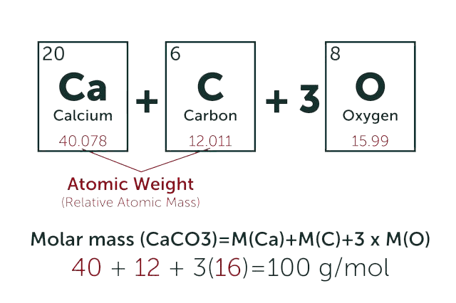
* Total Molecular Weight:
g/mol
Tip: Chemical formula is case sensitive. C22H30N4O √ c22h30n40 ╳