Chromomycin A2
* Please be kindly noted products are not for therapeutic use. We do not sell to patients.

| Category | Antibiotics |
| Catalog number | BBF-00639 |
| CAS | 6992-70-7 |
| Molecular Weight | 1211.30 |
| Molecular Formula | C59H86O26 |
| Purity | >98% by HPLC |
Online Inquiry
Description
Chromomycin A2 is produced by the strain of Streptomyces olivochromogenes 69895. A minor, more hydrophobic analogue of the chromomycin complex of the aureolic acid class; originally isolated from S. Aburaviensis and named aburamycin; exhibits a broad biological profile as an antibacterial, antifungal and antitumor agent.
Specification
| Synonyms | Aburamycin A; NSC 131187 |
| Storage | -20°C |
| IUPAC Name | [(2S,3S,4S,6S)-6-[(2R,3R,4R,6S)-6-[(2R,3R,4R,6S)-6-[[(2S,3S)-6-[(2S,4R,5S,6R)-5-acetyloxy-4-[(2R,4R,5R,6R)-4-hydroxy-5-methoxy-6-methyloxan-2-yl]oxy-6-methyloxan-2-yl]oxy-3-[(1S,3S,4R)-3,4-dihydroxy-1-methoxy-2-oxopentyl]-8,9-dihydroxy-7-methyl-1-oxo-3,4-dihydro-2H-anthracen-2-yl]oxy]-3-hydroxy-2-methyloxan-4-yl]oxy-3-hydroxy-2-methyloxan-4-yl]oxy-4-hydroxy-2,4-dimethyloxan-3-yl] 2-methylpropanoate |
| Canonical SMILES | CC1C(C(CC(O1)OC2C(CC3=C(C2=O)C(=C4C(=C3)C=C(C(=C4O)C)OC5CC(C(C(O5)C)OC(=O)C)OC6CC(C(C(O6)C)OC)O)O)C(C(=O)C(C(C)O)O)OC)OC7CC(C(C(O7)C)O)OC8CC(C(C(O8)C)OC(=O)C(C)C)(C)O)O |
| InChI | InChI=1S/C59H86O26/c1-22(2)58(70)85-57-29(9)78-43(21-59(57,11)71)82-37-18-40(74-25(5)49(37)66)81-36-19-42(75-26(6)48(36)65)84-56-33(55(73-13)52(69)47(64)24(4)60)15-31-14-32-16-35(23(3)46(63)44(32)50(67)45(31)51(56)68)80-41-20-38(54(28(8)77-41)79-30(10)61)83-39-17-34(62)53(72-12)27(7)76-39/h14,16,22,24-29,33-34,36-43,47-49,53-57,60,62-67,71H,15,17-21H2,1-13H3/t24-,25-,26-,27-,28-,29+,33+,34-,36-,37-,38-,39-,40+,41+,42+,43+,47+,48-,49-,53+,54+,55+,56+,57+,59+/m1/s1 |
| InChI Key | WPLCTUHONLQGIX-TWTCTLMISA-N |
| Source | Streptomyces sp. |
Properties
| Appearance | Yellow Powder |
| Antibiotic Activity Spectrum | fungi; tumor |
| Melting Point | 182-187 °C |
| Solubility | Soluble in ethanol, methanol, DMF or DMSO. Limited water solubility. |
Reference Reading
1. Chromomycin A2 induces autophagy in melanoma cells
Hozana Patrícia S Freitas, Diego Veras Wilke, Larissa Alves Guimarães, Danilo Damasceno Rocha, Jesús Martín, Otília Deusdênia Loiola Pessoa, Paula Christine Jimenez, Thiciana da Silva Sousa, Letícia Veras Costa-Lotufo, Fernando Reyes Mar Drugs . 2014 Dec 4;12(12):5839-55. doi: 10.3390/md12125839.
The present study highlights the biological effects of chromomycin A2 toward metastatic melanoma cells in culture. Besides chromomycin A2, chromomycin A3 and demethylchromomycin A2 were also identified from the extract derived from Streptomyces sp., recovered from Paracuru Beach, located in the northeast region of Brazil. The cytotoxic activity of chromomycin A2 was evaluated across a panel of human tumor cell lines, which found IC50 values in the nM-range for exposures of 48 and 72 h. MALME-3M, a metastatic melanoma cell line, showed the highest sensitivity to chromomycin A2 after 48h incubation, and was chosen as a model to investigate this potent cytotoxic effect. Treatment with chromomycin A2 at 30 nM reduced cell proliferation, but had no significant effect upon cell viability. Additionally, chromomycin A2 induced accumulation of cells in G0/G1 phase of the cell cycle, with consequent reduction of S and G2/M and unbalanced expression of cyclins. Chromomycin A2 treated cells depicted several cellular fragments resembling autophagosomes and increased expression of proteins LC3-A and LC3-B. Moreover, exposure to chromomycin A2 also induced the appearance of acidic vacuolar organelles in treated cells. These features combined are suggestive of the induction of autophagy promoted by chromomycin A2, a feature not previously described for chromomycins.
2. Tailoring modification of deoxysugars during biosynthesis of the antitumour drug chromomycin A by Streptomyces griseus ssp. griseus
Jürgen Rohr, Alfredo F Braña, José A Salas, Carmen Méndez, Nuria Menéndez, Mohammad Nur-E-Alam Mol Microbiol . 2004 Aug;53(3):903-15. doi: 10.1111/j.1365-2958.2004.04166.x.
Chromomycin A3 is a member of the aureolic acid group family of antitumour drugs. Three tailoring modification steps occur during its biosynthesis affecting the sugar moieties: two O-acetylations and one O-methylation. The 4-O-methylation in the 4-O-methyl-D-oliose moiety of the disaccharide chain is catalysed by the cmmMIII gene product. Inactivation of this gene generated a chromomycin-non-producing mutant that accumulated three unmethylated derivatives containing all sugars but differing in the acylation pattern. Two of these compounds were shown to be substrates of the methyltransferase as determined by their bioconversion into chromomycin A2 and A3 after feeding these compounds to a Streptomyces albus strain expressing the cmmMIII gene. The same single membrane-bound enzyme, encoded by the cmmA gene, is responsible for both acetyl transfer reactions, which convert a relatively inactive compound into the bioactive chromomycin A3. Insertional inactivation of this gene resulted in a mutant accumulating a dideacetylated chromomycin A3 derivative. This compound, lacking both acetyl groups, was converted in a two-step reaction via the 4E-monoacetylated intermediate into chromomycin A3 when fed to cultures of S. albus expressing the cmmA gene. This acetylation step would occur as the last step in chromomycin biosynthesis, being a very important event for self-protection of the producing organism. It would convert a molecule with low biological activity into an active one, in a reaction catalysed by an enzyme that is predicted to be located in the cell membrane.
3. [Chromomycin A(2) induces apoptosis of HepG2 cells in vitro]
Meijuan Xie, Zhenning DU, Yan Wang, Yuanyuan Lu, Weiwei Bao Nan Fang Yi Ke Da Xue Xue Bao . 2014 Oct;34(10):1449-53.
Objective:To study the effect of chromomycin A(2) in inducing apoptosis of HepG2 cells and explore the molecular mechanism.Methods:HepG2, MCF-7, A549, and 7901 cells were exposed to chromomycin A(2) and the changes in the cell viability were detected using MTT assay. The changes in the chromatins were observed with laser scanning confocal microscope after incubation of the cells with chromomycin A(2) (60 nmol/L) for 24 h. The changes in cell morphology were examined with a phase-contrast microscope, and the apoptotic cell populations, fluorescent intensity of reactive oxygen species (ROS) and mitochondrial membrane potential were determined using flow cytometry.Results:Chromomycin A(2) significantly inhibited the proliferation of the cells in a time- and dose-dependent manner, and caused changes in the cell morphology and cell apoptosis. Exposure of the cells to chromomycin A(2) resulted in chromatin condensation, ROS generation, and reduction of the mitochondrial membrane potential.Conclusion:Increased ROS and mitochondria damage may importantly contribute to chromomycin A(2)-induced apoptosis in HepG2 cells.
Recommended Products
| BBF-01825 | Loganin | Inquiry |
| BBF-05862 | Epirubicin | Inquiry |
| BBF-01693 | Doxorubicin EP Impurity A (Daunorubicin) | Inquiry |
| BBF-03755 | Actinomycin D | Inquiry |
| BBF-02614 | Nystatin | Inquiry |
| BBF-01829 | Deoxynojirimycin | Inquiry |
Bio Calculators
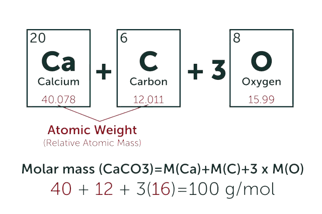
* Our calculator is based on the following equation:
Concentration (start) x Volume (start) = Concentration (final) x Volume (final)
It is commonly abbreviated as: C1V1 = C2V2
* Total Molecular Weight:
g/mol
Tip: Chemical formula is case sensitive. C22H30N4O √ c22h30n40 ╳